30+ Vibrate Cochlea Diagram PNG. The cell bodies of the cochlear nerve (spiral ganglion) that transmit information from different regions of the basilar membrane therefore encode frequency tonotopically. Investigated ultimately in living cochleae.
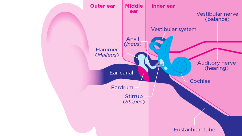
How the ear works - Action on Hearing Loss from actiononhearingloss.org.uk The vibration of the stapes is transferred into the cochlea by way of the oval window, and fluids within the scala vestibuli and scala tympani begin to move. In acoustics, the decibel is quantified. This occurs at the organ of corti, a structure.
Cochlea в национальной медицинской библиотеке сша по медицинским предметным рубрикам (mesh).
The ossicle bone known as the. Learn vocabulary, terms and more with flashcards, games and other study tools. Cochleae) is part of the inner ear osseous labyrinth found in the petrous temporal bone. The cochlea not only acts as a passive detector of sound, however, but can also produce tones itself.
Bagikan Artikel ini
Belum ada Komentar untuk "Vibrate Cochlea Diagram"

Belum ada Komentar untuk "Vibrate Cochlea Diagram"
Posting Komentar